Deformities - Injuries
Planovalgus deformity
The deformity appears in people otherwise healthy at the 4th or 5th decade of life. Pain usually precedes for months on the medial aspect of the foot just beneath the medial malleolus (ankle area). This is actually a tendinitis of the posterior tibialis tendon which is gradually frayed while the heel turns laterally and the plantar arch is progressively lost. This situation makes walking painful and difficult.
Various supportive insoles may be used in early stages with limited results. Surgery aims at restoring the anatomical axis of the foot. Surgical techniques are combinations of tendon transfers, osteotomies, arthrodesis. Age, weight, degree of deformity, any concomitant arthritis of the adjacent joints will all help surgeon to choose the method that will be used in a given patient.
Surgery is done under general or spinal anaesthesia. The patient is discharged from the hospital after a few days. Plaster or some cast can be applied for a few weeks postoperatively and weight bearing is individualized. Stitches are removed 2 weeks postoperatively.
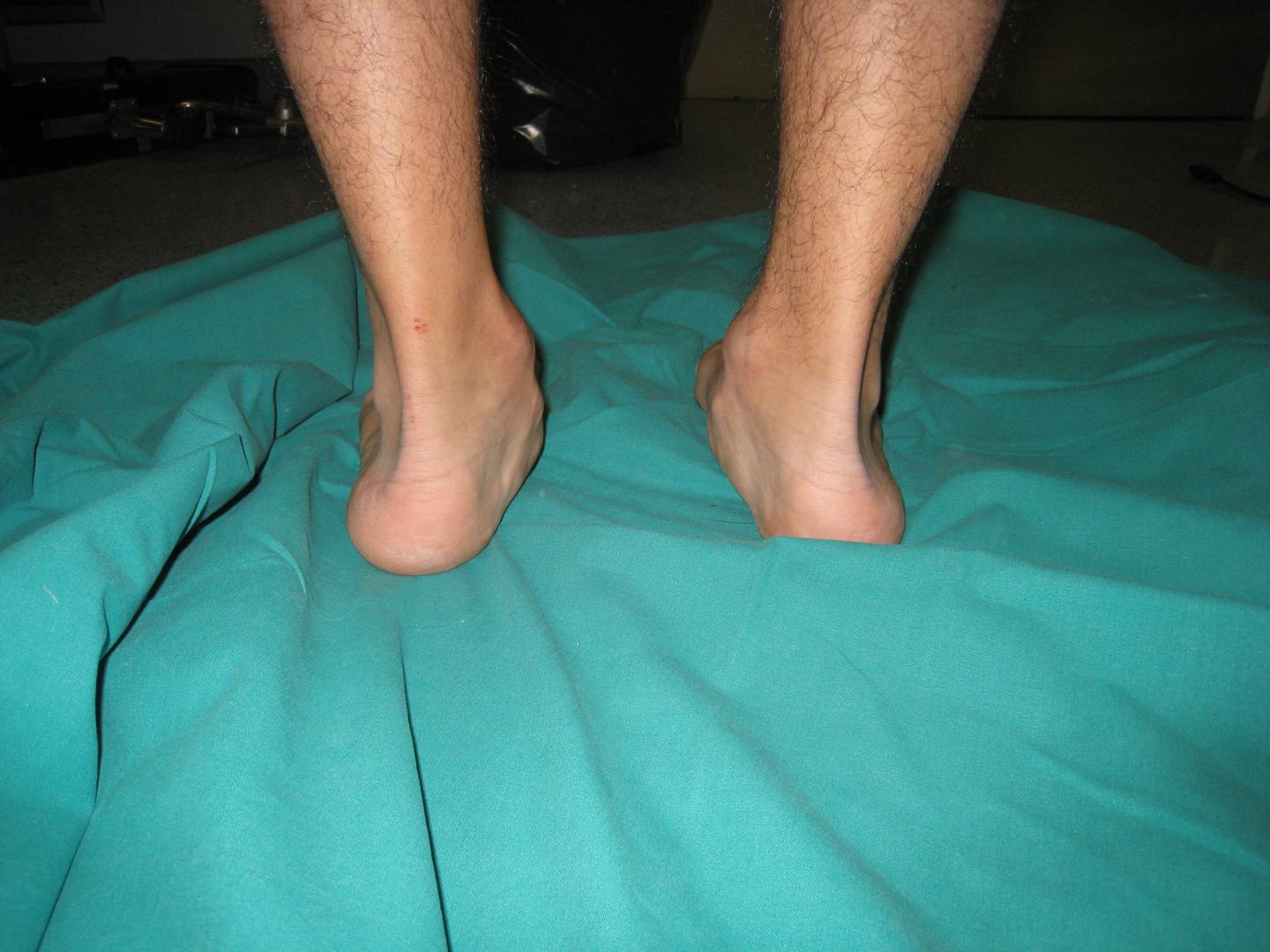
Αμφοτερόπλευρη επίκτητη βλαισοπλατυποδία προεγχειρηγτικά
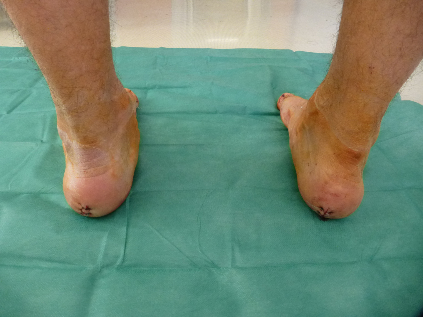
Αμφοτερόπλευρη επίκτητη βλαισοπλατυποδία μετεγχειρητικά